Sinus CT Scans
Wellington S. Tichenor, M. D.
New York, New York
(212)517-6611
If you haven't seen the rest of this website, please look at the rest of it.
There is a lot of valuable information in this site.
Because the images below are large files, sometimes they may be difficult to print.

The following is designed to enable you to develop a basic understanding of sinus anatomy as well as CT scans, both normal and abnormal. We will review several CT scans, but start with a drawing to start to orient you.
There are four sets of sinuses: maxillary, ethmoid, frontal and sphenoid sinuses. We will examine most of them in the following series of drawings and CT scans. The initial concepts are a little difficult to understand, but will become clearer when we get to the CT scans.


LEGEND:
F - Frontal sinuses,
E - Ethmoid sinuses,
M - Maxillary sinuses,
O - Maxillary sinus ostium,
SS - Sphenoid sinus
ST- Superior turbinate,
T - Middle turbinate,
IT- Inferior turbinate,
SM- Superior meatus,
MM- Middle meatus,
SR - Sphenoethmoidal recess,
S- Septum,
ET - Eustachian tube orifice,
A - Adenoids
. Courtesy of Astra Pharmaceuticals
In the first graphic representation, the three overlapping flaps of tissue, called turbinates (inferior - IT, middle - T, and superior - ST ) protect the openings of the sinuses, and allow humidification, filtration and warming of air. The frontal (F) sinus is seen in this view, but is not usually involved to any great extent in sinusitis. The sphenoid sinus (SS) is also seen in this view, and is sometimes involved in sinusitis. The sphenoid sinus drains into the sphenoethmoidal recess (SR)
In the second graphic representation, the maxillary sinuses (M) drain through the maxillary sinus ostia (O) into the middle meatus (MM). It should be noted that in this graphic diagram, the opening at O appears to be extremely large. In actuality, it is the size of a pin head and actually follows a rather circuitous route as you will see on the CT scans which follow. The ethmoid sinuses (E) drain into both the middle meatus as well as into the superior meatus (SM).
The middle meatus (MM) is bounded by the middle turbinate (T) and the inferior turbinate (IT). (There is also a superior turbinate (ST), but that is relatively unimportant.)
Another important structure is the "ostiomeatal unit" which is the outflow tract from the sinuses and includes the ostium of each sinus as well as the meati. When blocked, the ostiomeatal unit can cause obstruction of the sinuses, analogous to putting a plug in a bathtub.
The frontal sinuses (F) are occasionally important, but will not be dealt with to any great extent in this discussion. The septum (S) creates a barrier between the two sides of the nose. If it is deviated to a great enough extent, an obstruction can occur. Occasionally, there may be a perforation (hole) in the septum, which can cause problems with the architectural support of the nose.
The first CT scan is relatively normal:

LEGEND:
+ - border of maxillary sinus,
* - maxillary sinus ostium,
U - uncinate process,
E - ethmoid sinuses,
IT- inferior turbinate,
MT- middle turbinate,
S - septum,
C - concha bullosa.
Note that the CT scan is a computerized X-Ray taken in the same way as the first diagram is drawn, as if you were able to look head on into the sinuses. Note that the patient's right side is on your left as indicated by the "right" mark in the upper left hand corner.
On the CT scan, bone appears white, air appears black, and soft tissue, fluid, or muscle is varying shades of gray. Of note in the bottom portion of the scan is a ray pattern emanating from the teeth. This is as a result of poor penetration of the x-rays through the metal in the teeth.
When we evaluate the sinuses for sinusitis, we look for thickening of the lining of the sinus. Note on the patient's left side (your right) at the + sign, there is a very sharp distinct border between the black air in the maxillary sinus and the white bone. As you will see later, sinusitis is manifested by grayish thickening of the lining of the sinus.
The asterisk (*) is at the point where drainage occurs from the maxillary sinus into the nose through part of the ostiomeatal unit. The maxillary sinus ostia is bounded below by the uncinate process (U) and above by the lower bony portion of the ethmoid sinuses (E). A narrowing in this area obviously can be very critical. The ethmoid sinuses, as can be seen, are much smaller than the maxillary sinuses.
On the right side (your left), one can see the middle turbinate (MT) as well as inferior turbinate (IT). There is a slight deviation of the septum (S) to the right side (your left), but in this case it is unlikely that it is causing any obstruction. Of note is that there is air contained in the middle turbinate on the left (C-short for concha bullosa). This represents a normal anatomical variant in which the ethmoid sinuses have pushed down into the middle turbinate. In this case, it does not appear to have caused a problem, but often it will cause a significant enlargement of the middle turbinate and consequently an obstruction on one side of the nose.
The next CT scan is from a patient with significant sinus disease.

LEGEND:
M - maxillary sinus,
+ - thickening of the maxillary sinus,
E - ethmoid sinuses,
P - polyp,
O - maxillary sinus ostium,
* - middle meatus.
Attention should first be directed to the + sign on the right side. Compared to the previous scan, there is a significant amount of grayish thickening between the white bone and the black sinuses. Any thickening over 3 mm is definitely abnormal. Note that this thickening involves almost the entire maxillary sinus on both sides, but more so on the right side.
Compare the area on the right side where the maxillary sinus ostium (O) was observed on the previous CT scan. There is no opening now, only the gray tissue completely blocking the ostium. Not surprisingly, this patient had a great deal of pain as a result of that blockage. Although there is more thickening of the sinus lining on the right, there is more room to breathe through the nose on the right side (*) than on the left side. This is largely as a result of the deformity of the middle turbinate, located just above the asterisk (compare to the opposite side). This may have contributed to the sinus disease in this case, causing obstruction of the ostiomeatal unit. Not surprisingly the ethmoid sinuses were involved as well. The ethmoid sinuses are either filled with polyps (P) or the lining is thickened. There is very little air left in the ethmoid sinuses (E).
This X-Ray is from another patient with fairly severe sinus disease:
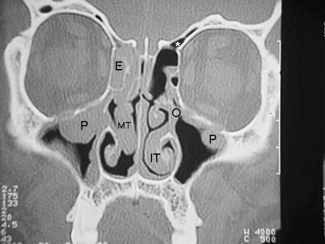
LEGEND:
MT- middle turbinate,
IT- inferior turbinate,
P - polyp or cyst,
E - ethmoid sinuses,
O - maxillary sinus ostium,
* - frontal sinus.
As compared to the previous x-ray, this patient has larger polyps or cysts (P) (it is often difficult to tell the difference on CT scan) on the right side, with significant obstruction of the ostium. On the left side the ostium (O) cannot be clearly seen, but you get the idea of where it's supposed to be. The ethmoid sinuses (E) on the right side are completely obstructed, being filled with either polyps, cysts or thickening of the sinuses. On the left side, there is some thickening, but as you can see, for the most part the ethmoid sinuses are fairly clear. The asterisk on the left side represents the point at which the ethmoid sinuses merge into the frontal sinuses. Note the difference in size between the middle turbinate (MT) on the right and left side, and also the inferior turbinates (IT) on each side.
This CT scan was done after surgery was performed on the patient
whose CT scan you just reviewed.

LEGEND: E - Ethmoid sinuses,
O - maxillary sinus ostium,
M - maxillary sinuses,
S - septum,
MT - middle turbinate,
IT - inferior turbinate.
That's pretty impressive isn't it ?, even if you're a lay-person. As you can see, the polyps that were previously in the maxillary sinus (M) are now gone and the opening at the maxillary sinus ostia (O) is wide open, having had the uncinate process removed. The ethmoid sinuses (E) on both sides have been cleaned out. The surgery which was performed did not involve extensive removal of the lining of the sinuses. Just opening them up and allowing them to "breathe" is often enough to prevent severe disease. Of note, however, is the fact that this patient still doesn't have a normal nasal airway. (Compare to the first CT scan. Note the size of the black area next to the middle turbinate (MT) and inferior turbinate (IT).)
In addition to having sinus problems, this patient also has allergy problems which had to be treated in order to prevent future nasal problems and sinus disease.
We hope that the short course on X-Rays of the sinuses has been helpful in understand a little more about sinusitis.
We suggest going to
Technical Information for Physicians or
Going to the Allergy Page
as 70-80% of sinusitis sufferers have allergy problems
(including a lot that don't realize it).
Any comments on this site would be appreciated. Please send mail
to wtichenor@sinuses.com. We will attempt to answer as many
questions as is feasible personally, but are obviously limited
due to time constraints. Any items of general interest will be
included within the website.
The contents of sinuses.com © 1997-2010 by Wellington S. Tichenor,
M.D. Last updated March 14, 2010.Reproduction for educational, not-for-profit purposes is
permitted if this source is credited and the author of this
website is notified of any reproduction for other than personal
use. If used on the internet, a link would be appreciated.
|Welcome Page|Table of Contents|Go to Top of Page|Allergy Page|Asthma Page|E-Mail